

Introduction to Medical Imaging
Medical Imaging has revolutionized the healthcare field, allowing doctors to see inside the human body with remarkable detail and precision. One of the most critical advancements in medical imaging technology is the development of computed tomography (CT) scans. CT scans have become indispensable for diagnosing various medical conditions and guiding treatment plans. In this blog post, we will explore how CT technology has evolved, mainly focusing on the exciting advancements of 3D and 4D Imaging. Get ready to dive into a world where images come alive, revealing secrets once hidden beneath our skin!
What is a CT Scan?
A CT scan, or computed tomography scan, is a medical imaging technique that uses X-ray technology to create detailed cross-sectional images of the body. It provides healthcare professionals valuable information about internal structures and can help diagnose various conditions.
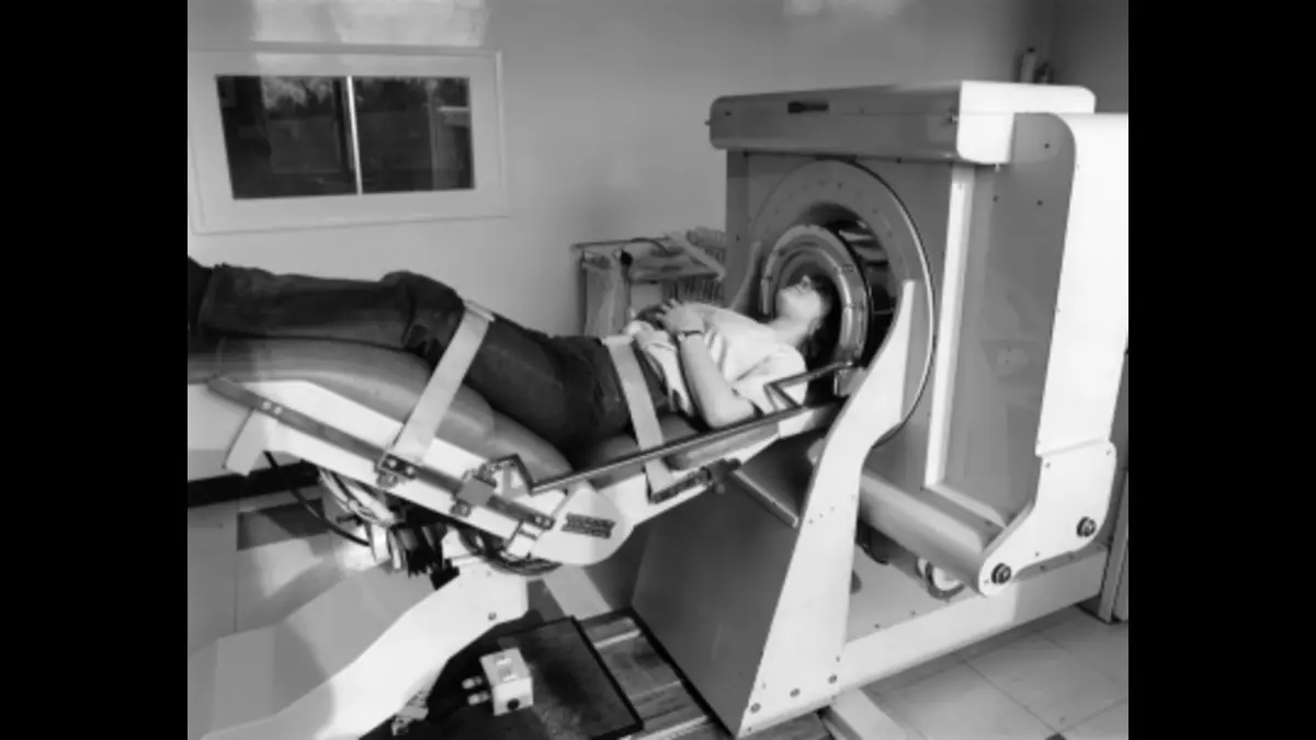
During a CT scan, the patient lies on a table that moves through a doughnut-shaped machine called the gantry. The gantry houses an X-ray tube and detector array that rotate around the body, capturing multiple images from different angles. These images are then processed by computer software to produce detailed cross-sectional slices.
One of the critical advantages of CT scans is their ability to capture images in axial, coronal, and sagittal planes. This enables doctors to view anatomical structures from different perspectives and detect abnormalities that may not be apparent in traditional two-dimensional X-rays.
A Brief History of CT Technology
Input TomtomographyT) technology has made significant strides since its inception. Medical Imaging began in the early 1970s when British engineer Godfrey Hounsfield developed the first CT scanner. His groundbreaking work earned him a Nobel Prize in Physiology or Medicine.
The initial CT scanners were bulky and slow compared to today’s sleek and efficient machines. They used X-ray beams that rotated around the patient to capture multiple cross-sectional images, which were then reconstructed by computer algorithms. This revolutionized diagnostic medicine by providing detailed views of internal organs and structures that were previously inaccessible.
Over time, advancements in CT technology have allowed for faster scanning times, improved image quality, and reduced radiation exposure—the introduction of spiral/helical scanning in the late 1980s enhanced image acquisition speed and efficiency.
Advancements in CT Tech 3D and 4D Imaging
Medical Imaging has come a long way since its inception, with continuous advancements revolutionizing the field. One of the most notable developments is the introduction of 3D and 4D Imaging in CT technology.
Gone are the days when traditional two-dimensional images were sufficient for diagnosis. With the advent of 3D and 4D Imaging, healthcare professionals can now obtain a more comprehensive view of internal structures.
In three-dimensional Imaging, multiple X-ray projections are compiled to create a detailed representation of an organ or body part. This allows physicians to visualize complex anatomical structures from various angles, aiding in accurate diagnoses and treatment planning.
Benefits of 3D and 4D Imaging in Medical Diagnosis
Advancements in CT technology have revolutionized medical Imaging, mainly by introducing 3D and 4D Imaging. These cutting-edge techniques have significantly enhanced the accuracy and precision of medical diagnosis.
One of the key benefits of 3D and 4D Imaging is their ability to provide a more detailed view of internal structures within the body. Unlike traditional 2D images, which can sometimes be limited in their ability to show depth or spatial relationships, these advanced imaging techniques create a three-dimensional representation that allows healthcare professionals to understand complex anatomical features better.
The Future of CT Scans: Artificial Intelligence and Machine Learning
As technology continues to advance at an unprecedented pace, the field of medical Imaging is also undergoing a revolutionary transformation. One exciting area of development in CT scan technology is the integration of artificial intelligence (AI) and machine learning algorithms.
Healthcare professionals can expect more accurate diagnostics and improved patient outcomes with AI-powered CT scans. By leveraging vast amounts of data, machine learning algorithms can quickly analyze images and assist radiologists in detecting abnormalities that may have been missed by the human eye alone.
Limitations and Challenges of CT Technology
While CT technology has revolutionized medical Imaging, it is not without its limitations and challenges. One major limitation is the exposure to ionizing radiation during a CT scan. Although the amount of radiation used in modern CT scanners has significantly decreased, repeated exposure can still pose risks to patients.
Another challenge is the potential for false positives or false negatives in interpretation. Even with advancements in image resolution and clarity, there can still be instances where abnormalities are missed or typical structures are mistaken for abnormalities. This highlights the importance of skilled radiologists who can accurately interpret these images.
Conclusion: The Ongoing Evolution of Medical Imaging
As we have seen throughout this article, the advancements in CT technology have revolutionized medical Imaging and greatly enhanced our ability to diagnose and treat various conditions. From its humble beginnings as a simple two-dimensional scan, CT scanning has evolved into a powerful tool that can provide detailed 3D and even 4D images of the human body.
With the introduction of 3D and 4D Imaging, doctors now have access to more information than ever. These advanced imaging techniques allow for better visualization of complex structures, improved accuracy in detecting abnormalities, and enhanced surgical planning. They have become invaluable in diagnosing conditions such as cancer, cardiovascular diseases, neurological disorders, and much more.


